


Ophthalmology Surgery 2018
Event begins in
Ophthalmology Conference Highlights
About Conference
EuroSciCon will be holding a conference on Ophthalmology Surgery 2018 from July 30-Aug 1st, 2018 in Barcelona, Spain. The theme of this year’s meeting is “Vision for a better life and novel concepts in Ophthalmology Surgery” which will provide an international platform for discussion of present and future challenges in primary care, patient care, continuing education and expertise meeting. Ophthalmology Surgery conferences aim to promote a forum for international researchers from various areas of Ophthalmology, Optometry, Life sciences and allied groups by providing a platform for critical analysis of new data, and to share novel research findings and results about all aspects regarding advances in various branches of Ophthalmology Surgery. We are awaiting a great scientific faculty from USA, Europe as well as other continents and expect a highly interesting scientific as well as a representative event.
What’s New
Ophthalmology Surgery 2018 includes international attendee workshops, lectures and symposia, including a designated registration area, a refreshment break and gala lunch. Ophthalmology Surgery conferences can join the EuroSciCon as an international member to receive discounts on registration. So come and join leading experts and allied professionals from July 30-Aug 1st, 2018 in Barcelona, Spain to keep up with the rapidly accelerating pace of change that is already having an impact on the field of Ophthalmology Surgery and will continue to in the future.
Session and Tracks
SESSION AND TRACKS
Corrective Surgery: Corrective laser eye surgery is by far the most popular eye surgery in today’s times. It has taken the world by storm, with approximately 2,000 people having it performed every day. The two most common methods of corrective surgery are LASIK (Laser-Assisted In Situ Keratomileusis) and PRK (photorefractive keratectomy). Laser eye surgery can correct myopia, hyperopia, and astigmatism, thereby eliminating an individual’s dependency on corrective lenses.
o Most popular, LASIK uses a precision laser to remove corneal tissue and reshape the cornea to change its level of focus.
o PRK is preferred for individuals with thin corneas where creating a flap may be more difficult.
Both types of corrective surgery take approximately 15 minutes per eye. New “bladeless” wave front technology has been introduced to mitigate some of the issues caused by aberrations in flap edges.
Cataract Surgery: Cataracts, commonly affecting both eyes, occurs in over half of adults over 60 years. The surgery to remove cataracts is a very safe and successful procedure, with over 1.5 million cataract surgeries performed in the United States to date. It is often recommended for those individuals who have vision loss that is impeding their daily life, but may be avoided for those who have additional eye diseases. Cataract surgery is performed with minimal sedation and generally takes less than 30 minutes. During surgery, an artificial lens is put in place of the original to restore vision.
There are three different approaches to cataract surgery:
Phacoemulsification
Most common, this approach requires only local anesthesia. Ultrasonic vibrations dissolve the cloudy lens via an inserted probe.
Extra capsular cataract surgery
This type of surgery is often performed when the lens is too dense for phacoemulsification. This surgery requires sutures, and recovery is longer.
Intra capsular cataract surgery
This technique requires a larger incision, and the entire lens and surrounding capsule are removed. This method is rare, and is only used when existing eye trauma makes it the most practical option.
Over time, the lens replacement may begin to cloud. This is easily corrected with an additional laser treatment.
Glaucoma Surgery: Glaucoma results in raised intraocular pressure and vision loss over time. Unfortunately, surgery for glaucoma cannot reverse this vision loss. However, surgery can reduce the intraocular pressure when medication is not a sufficient solution. If necessary, glaucoma surgery can be performed multiple times with low risk.
There are two kind of common glaucoma surgery: laser and conventional.
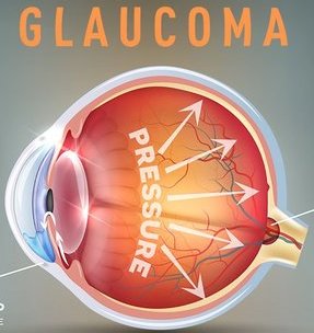
Laser Surgery – During laser surgery, a laser is used to make a small opening in the eye’s drainage system to help increase fluid drainage out of the eye. When laser surgery unable to solve the condition & condition re-emerges, the ophthalmologist may refer for conventional surgery.
Conventional Surgery – During conventional surgery, a new drainage canal is created that allows intraocular fluid to flow out of the eye.
Lid Repair Surgery: Lid repair may be shown for both cosmetic and functional reasons. Lid repair is most generally used to treat ptosis, also called as droopy eyelids. This has become an extremely famous surgery as the condition can cosmetically age the patient, obstruct vision, or make a visual incongruence between the eyelids.
Surgeries for ptosis can incorporate blepharoplasty (ptosis resulting from aging), Fasanella-Servat and Müller’s muscle-conjunctival resection (for mild to moderate ptosis), and frontalis suspension (for more extreme congenital cases).
Orbital Surgery: Orbital eye surgery is demonstrated for conditions for example anopthalmia, enucleation, and evisceration. These types of surgeries are indicated for individuals with: congenital defects resulting in absent eyes, large eye tumors, or when the eye cannot be preserved due to trauma.
Surgical methodologies incorporate introducing an orbital implant for example the Orbital Tissue Expander, and bioceramic or silicone orbital implants. The Orbital Tissue Expander helps the preserve the congruency of facial structure in congenital cases where maintaining pressure on the surrounding structures is imperative to proper growth.
Orbital implants are utilized to protect the regular structure of the eye orbit and provide support for the placement of an artificial eye. Recent advancements in orbital implants have aided in improving implant motility and allowing for the natural movement of artificial eyes. Stable materials that are biocompatible, bioinert, nontoxic, and non-allergenic provide a lower rate of migration and rejection in patients.
Eye muscle surgery (strabismus) : Strabismus is a vision problem in which both eyes do not focus at the same object at the same time. Strabismus most often begins in early childhood. It is sometimes called crossed-eyes, walleye, or squint.
Normally, the muscles attached to each eye work together to move both eyes in the same direction at the same time. Strabismus occurs when the eye muscles don't work accurately to control eye movement. When the eye muscles don't work as they should, the eyes may become misaligned and the brain may not be able to merge what one eye sees with what the other eye sees.
The most common treatments for strabismus are:
Glasses. Wearing glasses sometimes correct mild strabismus.
A temporary eye patch over the stronger eye.This can make the weak eye stronger, which may help in aligning the eyes. Your child may have to wear the patch some or all of the time for a few weeks or months.
Surgery on the eye muscles. This is the only way to improve vision and for betterment in alignment of the eyes. It may take more than one surgery, and your child may still need to wear glasses.
Vitrectomy / Vitreo-retinal surgery : Vitrectomy is a surgery to remove the vitreous gel from the center of the eye. It may be done when there is a retinal detachment or if blood in the vitreous gel (vitreous discharge) does not clear on its own. Removing the vitreous gel gives your eye specialist better access to the back of the eye. Vitrectomy is done by an eye doctor (ophthalmologist) who has special training in treating problems of the retina.
During surgery, the doctor uses small devices to remove the vitreous gel. Then the specialist may treat other eye issues, for example a retinal detachment, vitreous drain, scar tissue on the retina, or tears or holes in the macula.
At the end of the surgery, the specialist may inject an oil or gas bubble into the eye. This gently presses the retina against the wall of the eye. If an oil bubble is used, the doctor will need to evacuate/remove the oil after the eye has healed.
Refractive surgery: Refractive surgery is used to correct common eye disorders such as nearsightedness, farsightedness, and astigmatism. Refractive surgery is designed to alter the shape of the cornea in order to improve the patient’s vision.
For people who are myopia, some refractive surgery techniques will minimize the curvature of a cornea that is too steep so that the eye's focusing power is lessened. Images that are focused in front of the retina, due to a longer eye or steep corneal curve, are pushed closer to or directly onto the retina following surgery.
Hyperopia people will have refractive surgery procedures that achieve a steeper cornea to increase the eye's focusing power. Images that are focused beyond the retina, due to a short eye or flat cornea, will be pulled closer to or directly onto the retina after surgery.
Astigmatism can be treated with refractive surgery techniques that selectively reshape part of an irregular cornea to make it smooth and symmetrical. The result is that images focus clearly on the retina rather than being distorted due to light scattering through an irregularly shaped cornea.
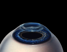
Corneal surgery/ corneal grafting/ corneal transplant: The cornea is the clear outer lens on the front of the eye. The cornea is made of layers of cells. These layers work together to protect your eye and provide clear vision.
Your cornea must be clear, smooth and healthy for good vision. If it is swollen, scarred or damaged, light is not focused accurately into the eye. As a result, your vision is blurry or you see glare. A cornea transplant replaces diseased or scarred corneal tissue with healthy tissue from an organ donor.
There are two main kinds of corneal transplants: traditional, full thickness cornea transplant (also known as penetrating keratoplasty, or PK) and back layer/ Partial-thickness cornea transplant (also known as endothelial keratoplasty, or EK).
A graft replaces central corneal tissue, damaged due to disease or eye injury, with healthy corneal tissue donated from a local eye bank. An affected cornea affects your vision by scattering or distorting light and causing glare and blurred vision. A cornea transplant may be necessary to restore your functional vision.
Corneal eye disease is the fourth most common cause of blindness (after cataracts, glaucoma and age-related macular degeneration) and affects more than 10 million people worldwide.
Oculoplastic Surgeries: Oculoplastic surgeons are ophthalmologists (eye doctors) who have specialized in eyelid and facial plastic surgery. Oculoplastic surgery is a general term used to represent a variety of procedures that involve the orbit, eyelids, & tear ducts. Ocular reconstructive surgery, aesthetic eyelid surgery, facial plastic surgery, and cosmetic procedures fall into this category.
Some types of oculoplastic surgery are considered both medically necessary and cosmetic. For instance, certain eyelid and periocular issues can affect a person’s appearance as well as their vision, eye comfort, and eye health.
Retinal detachment: Retinal separation/detachment is a serious condition of the eye in which the retina stops receiving oxygen. Your retina is the part of your eye that sends images through your optic nerve to the brain. Your retina contains millions of cells that detect light like a camera. It is part of the very back of your eyeball and is essential to your vision.
Retinal detachment occurs when the retina pulls away from the back of the eye and the blood supply. Without a blood supply, the retinal cells will start to die. This can make changeless harm to your vision. If the macula begins to loosen, your vision may be permanently damaged. If macula completely detaches, you may lose your vision entirely. Reattaching the retina quickly is essential to prevent such a serious complication.
Retinal detachment happens because the vitreous fluid of the eye drawn back from the back of the eye, pulling the retina and tearing it. Some causes and risk factors of retinal detachment include glaucoma, severe trauma, nearsightedness, previous cataract surgery, previous retinal detachment in your other eye, or family history of retinal detachment. Symptoms of a retinal detachment include frightening. Objects might appear to float in front of your eye, or a gray veil may move across your field of vision. If not treated quickly, a retinal detachment can cause you to lose your vision. Retinal detachment repair is a surgery used to restore circulation to the retina and restore vision.
Advanced Imaging technologies/Ophthalmology Imaging:
Advanced diagnostic and therapeutic decisions reside on the outcomes of imaging technologies have become a common practice in every ophthalmic subspecialty. New devices and tools for evaluating the retina and optic nerve head, such as scanning laser polarimetry and spectral-domain optical coherence tomography (OCT), are widely used in clinical practice. These technologies provide objective quantitative measurements and in vivo real-time images of ocular structures. The performance of imaging devices is continuously being improved, and thus knowledge of their applications, advantages, and limitations must also be continuously updated to optimize their management by clinicians. A spectral-domain optical coherence tomography (SD-OCT) device in pediatric ophthalmology practice, this mode of imaging has become a standard diagnostic tool. Also known as fourier-domain OCT, the technology has a much higher quality of image than the previous standard of care, time-domain OCT. These new machines scan 65 or more times faster than the older OCT images.
Macular Degeneration: Macular Degeneration is the most common cause of vision loss, affecting more than 10 million Americans – more than cataracts and glaucoma combined.
At present, Macular Degeneration is considered an incurable eye disease.
The macula is made up of millions of light-sensing cells that provide sharp, central vision. Macular Degeneration happened due to the damage of the central portion of the retina, the inside back layer of the eye that records the images we see and sends them via the optic nerve from the eye to the brain, and it controls our ability to read, drive a car, recognize faces or colors, and see objects in fine detail.
There are two common types of Macular Degeneration: “dry” and “wet.” Approximately 85% to 90% of the cases of Macular Degeneration are the “dry” (atrophic) type, while 10-15% is the “wet” (exudative) type.
Stages of Macular Degeneration
There are three steps of Age-related Macular Degeneration (AMD).
Early AMD – Most population do not experience vision loss in the early stage of AMD, which is why regular eye exams are important, particularly if you have more than one risk factor (see below). Early AMD is diagnosed by the presence of medium-sized drusen (yellow deposits underneath the retina).
Intermediate AMD – At this stage, there may be little vision loss, but there still may not be noticeable symptoms. A comprehensive eye exam with specific tests will look for larger drusen and/or pigment changes in the retina.
Late AMD – At this stage, vision loss has become noticeable.
Diabetic eye disease: If you have diabetes, your blood glucose, or blood sugar, levels are too high. People with diabetes can have an eye disease called diabetic retinopathy. This is when high blood sugar levels cause damage to blood vessels in the retina. These blood vessels can swell and leak. Or they can close, stopping blood from passing through. Over time, this can damage your eyes. Diabetic eye disease incorporates a group of eye problems that patient with diabetes may face as a complication of diabetes. People with diabetes are at risk for diabetic retinopathy, cataract and glaucoma. Diabetic eye disease has no warning signs. Finding and treating the disease earliest, before it effects vision impairments or blindness, is the best way to control diabetic eye disease. If you have diabetes, make sure you get a dilated eye examination at least once a year.
Diabetic retinopathy is the broadest diabetic eye disease and a main cause of blindness in adults. It is caused by changes in the blood vessels of the retina. In some people with diabetic retinopathy, blood vessels may swell and leak fluid. In other people, abnormal new blood vessels grow on the surface of the retina. A healthy retina is required for good vision. If you have diabetic retinopathy, at first you may not notice changes to your vision. But over time, diabetic retinopathy can get worse and cause vision loss. Diabetic retinopathy usually affects both eyes.
Ophthalmic oncology: Tumors can arise in almost any part of the eye. Some eye tumors can be quite serious, while others need no treatment.
Ocular Surface Tumors, Tumors may occur on the surface of the eye due to overexposure to the sun and other causes.
Uveal Melanoma, Melanoma of the uveal tract is an uncommon, but serious, condition that requires careful diagnosis and follow-up.
Orbital Tumors and Inflammation, The orbit is comprised of the bones of the eye socket, the eyeball, the eye muscles, the optic nerve and the surrounding fat. Any of these structures may form a tumor in adults and children.
Retinoblastoma, Tumors of the retina, in the back of the eye, are uncommon and are mostly found in young children, although they can develop at any age.
Ocular tumors including intraocular tumor resection, as well as laser, radiation therapy, and chemotherapy treatments for the eye, eyelid, orbit and conjunctiva, as needed.
Ocular injuries/ Eye Trauma: The structure of your face shields your eyes from injury. Still, wounds can damage your eye, sometimes severely enough that you could lose your vision. Most eye wounds are preventable.
Injuries to the eye and encompassing structures can be caused by blunt traumafrom sport balls, fists, or airsoft/pellet/paintball guns; sharp trauma for example a stick, knives; or chemical trauma for example splash from a caustic substance like a cleaning material or pool supplies. Safety glasses should be worn at all times while playing with airsoft/pellet/paintball guns and these should never be pointed at anybody faces.
The broadest type of injury occurs when something teases the outer surface of your eye. Certain jobs such as industrial jobs or hobbies such as carpentry make this type of injury more likely. It's also more likely if you wear contact lenses. Chemicals or heat can burn your eyes.
Ptosis is when the upper eyelid droops over the eye. “Ptosis” is the medical term for a drooping upper eyelid. Eyelid drooping can sometimes affect your vision if it’s severe. The eyelid may droop just a little, or so much that it covers the pupil. Ptosis can decrease or even block normal vision. Children and adults can have ptosis. Fortunately, this condition can be treated to improve vision as well as appearance.
You can get it several ways. Sometimes, babies are born with it. You could get ptosis as an adult when the nerves that control your eyelid muscles are damaged. It might follow an injury that loosen/weakens the muscles and ligaments that raise your eyelids. Sometimes, it comes with age. The skin and muscles around your eyes get weaker. Surgery- like LASIK or cataract surgery can stretch your eyelid. An eye tumor can cause ptosis, too.
Myasthenia gravis (MG) is a neuromuscular disease that leads to varying degrees of skeletal muscle weakness. The most commonly affected muscles are those of the eyes, face, and swallowing. It can result in double vision, drooping eyelids, trouble talking, and trouble walking. MG is an autoimmune disorder which results from antibodies that block or destroy nicotinic acetylcholine receptors at the junction between the nerve and muscle. This prevents nerve impulses from triggering muscle contractions.
Babies of mothers with myasthenia may have symptoms during their first few months of life, known as neonatal myasthenia. Diagnosis can be done by blood tests for specific antibodies, the edrophonium test, or nerve conduction studies.
Myasthenia gravis is generally treated with medications known as acetylcholinesterase inhibitors such as neostigmine and pyridostigmine. Immunosuppressant’s, such as prednisone or azathioprine, may also be used. The surgical removal of the thymus gland may improve the chances of success in certain cases.
Ophthalmic pathology focuses on diseases of the eye and its surrounding tissues. Precision diagnosis of diseases is provided by the ophthalmic pathology service. Diseased tissues are examined macroscopically, microscopically and on the ultra-structural level. Advanced genomic, proteomic, and cytogenetic techniques can be utilized to diagnose diseases at a molecular level. The pathologic diagnosis of the disease plays a vital role in patient care.
Thyroid eye disease (TED) is an eye condition in which the eye muscles and fatty tissue behind the eye become inflamed. This can cause the eyes to be pushed forward (‘staring’ or ‘bulging’ eyes) and the eyes and eyelids to become swollen and red. In some cases there is swelling and stiffness of the muscles that move the eyes so that the eyes are no longer in line with each other; this can cause double vision. In rare cases TED can cause blindness from pressure on the nerve at the back of the eye or ulcers forming on the front of the eyes.
Thyroid Eye Disease: Graves' eye disease or TED is an autoimmune disease. It occurs when the body’s immune system attacks the back of the eye and causes inflammation. It is mainly associated with an over-active thyroid due to Graves’ disease, although it does sometimes occur in people with an under-active or normally functioning thyroid. Graves’ disease is the most common cause of an over-active thyroid (hyperthyroidism) in the UK. Thyroid eye disease (TED) is an eye condition that causes the muscles and soft tissues in and around your eye socket to swell. It may also be called thyroid associated ophthalmopathy(TAO), thyroid orbitopathy, Graves’ orbitopathyor Graves’ ophthalmopathy (GO).
Unfortunately, many people with TED are left with a permanent change, usually in the physical appearance of their eyes or sometimes double vision. Surgery can improve appearance. In more severe cases it can be combined with other treatments such as steroids and radiotherapy. Rehabilitative surgery may involve:
'Decompression' surgery to create more space behind the eyes when there is pressure on the nerve or if there is a lot of protrusion of the eyeballs, in order to improve a person’s appearance
Eye muscle surgery to treat double vision;
Eyelid surgery to improve the appearance of the eyelids
Pediatric Ophthalmology Surgery: Whenever possible eye misalignment treated conservatively. In many children who are farsighted, crossing of the eyes can be treated with glasses in place of surgery. In many adults, symptoms of double vision can be controlled with prisms instead of surgery. Children born with ptosis have what is called congenital ptosis. This can be due to problems with the muscle that lifts the eyelid. However, there are some eye misalignment problems that conservative measures cannot help, and in these cases eye muscle surgery is necessary.
A pediatric ophthalmologist is an ophthalmologist who has training in order to better understand and serve the eyes and developing visual system of infants and children. The Pediatric Ophthalmology Service offers primary care of common children's eye problems as well as comprehensive evaluations, inpatient consultations, and follow-up for children with simple or complex eye and vision disorders. Children presenting with amblyopia, lacrimal disorders, strabismus, pediatric glaucoma, ocular cancers, inherited eye disease, and other eye conditions receive not only short-term treatment of the immediate problem, but also long-term rehabilitative care and monitoring of their visual development.
Tiny Robots Revolutionizing Eye Surgery: Machines that are able of making exact operations inside the human eye will make it conceivable to perform altogether new procedures. Tiny surgical robots "are revolutionizing eye surgery," said Simon Parkin at Technology Review. The Robotic Retinal Dissection Device, known as R2D2, enables specialists to produce miniscule incisions,
move membranes as small as a hundredth of a millimeter thick, and perform other staggeringly precise maneuvers on patients' eyes utilizing "a joystick and a camera feed."
Ocular surface diseases: Unlike the skin covering the rest of the body, the ocular surface is secured only by a thin layer of tear film. A steady tear film present when the eye is open is the key system to keep up the ocular surface healthy. Ocular surface disorder are disorders of the surface of the cornea—the transparent layer that forms the structure in front of the eye. These disorders incorporate dry eye syndrome, meibomian gland dysfunction, rosaceous, scarring from glaucoma medications, allergies, chemical burns, thermal burns, and immunological conditions such as Mucous Membrane Pemphigoid and Sjogren’s Syndrome.
Ocular surface diseases can extremely affect eyesight and quality of life. Symptoms may include blurry vision, discomfort or pain, redness and itching, and in severe cases, blindness due to corneal scarring.
Unfortunately, cases often go undiagnosed and undertreated, because of a lack of understanding of symptoms and inaccurate evaluation. And, as people are living longer, these disorders are becoming more prevalent.
Uveitis: Uveitis is swelling and irritation of the uvea. The uvea is the center layer of the eye. The uvea gives a large portion of the blood supply to the retina. Uveitis is a general term describing a gathering of inflammatory disorders that produces swelling and destroys eye tissues. These disorders can slightly decreased vision or lead to extreme vision loss.
The expression “uveitis” is utilized because the disorder often affects a part of the eye called the uvea. In any case, uveitis is not limited to the uvea. These diseases also affect the lens, retina, optic nerve, and vitreous, producing reduced vision or blindness. Uveitis may be occured by irritation or disorders occurring in the eye or it can be part of an inflammatory disease affecting other parts of the body. Uveitis can last for a short (acute) or a long (chronic) time. The severest forms of uveitis reoccur commonly.
Eye care professionals may depict the disorder more specifically as:
Anterior uveitis
Intermediate uveitis
Posterior uveitis
Panuveitis uveitis
Eye care professionals may also depict the disease as infectious or noninfectious uveitis.
Ocular inflammation: Ocular inflammation has become a hot topic in ophthalmology, involving also several areas of medicine: internal medicine, surgery, basic research, physiology, pharmacology, microbiology, immunology, rheumatology, pharmacology, or laboratory. There are many ocular inflammatory diseases in different locations, including orbit, ocular adnexa, ocular surface, conjunctiva, cornea, sclera, uvea, retinal vessels, and optic nerve.
Ocular inflammatory disease is an extremely common issue that challenges ophthalmologists in a number of ways. The ocular surface, made up of many important components, can suffer from a wide variety of inflammatory issues.
It can be caused by many different things; some of the most common include allergic, bacterial, chemical or viral conjunctivitis, blepharitis, keratitis sicca, sub-conjunctival hematomas and corneal abrasion. These patients need to be treated quickly and efficiently, because the small percentage of emergency visits resulting from much more serious ocular emergencies such as endophthalmitis and bacterial keratitis require more immediate and intensive care.
Market Analysis
The ophthalmic surgical instrument market to develop from $1.2 billion out of 2017 to $1.6 billion of every 2022, driven by 4 percent yearly development in worldwide ophthalmic surgeries. Expanding inclination for single utilize instruments and blades has driven these gadgets to represent almost 66% of worldwide market incomes. While surgical instruments are utilized as a part of a wide range of ophthalmic surgery, waterfall and retinal surgery represent almost 80 percent of incomes
Overall markets utilizing the accompanying geographic/financial classifications: the US, Western Europe, Japan, Other Wealthy Nations, China, India, Latin America, and Rest of World. This report incorporates advertise information for 2016, and we estimate showcase execution through 2022
Scope and Importance:
As per an examination it is assessed that around 48% of total populace is visually impaired due to waterfall and more than 60 million individuals experience the ill effects of glaucoma and it is evaluated that the number would ascend to 80 million before the finish of 2020. Maturing populace is the main source for refractive blunder issue in nations like the USA and Europe, it is seen that the refractive mistake issue is generally found in populace maturing over 40 years. After a colossal destruction of the market amid financial deplete in 2008-2009, ophthalmology advertise is making up for lost time to return to the ordinary as there is a fast development in populace bringing about expanded number of patients with eye maladies and the rate of waterfall and glaucoma are expanding each year
The Eye Care in Spain statistical surveying report :
Spain's market for ophthalmic gadgets is at present esteemed at USD XX billion for the year 2016 and is relied upon to achieve USD XX billion before the finish of 2021. The CAGR amid this time of the conjecture is anticipated to be XX%.
North America right now rules the worldwide market for ophthalmic gadgets. Developing countries are relied upon to develop at a decent pace inferable from the reality there is an expanded rate of waterfall in these regions
• Analysis of key supply-side and request patterns
• Detailed division of global and nearby items
• Historic volumes and qualities, organization and brand pieces of the pie
• Five year figures of market patterns and market development
• Robust and straightforward statistical surveying strategy, directed in-nation
• Analysis of key supply-side and request patterns
Learn More
Top Ophthalmology Universities:
Ophthalmology Universities in USA:
Johns Hopkins School of Medicine| UCSF School of Medicine| Perelman School of Medicine | Washington University| University of California San Diego| EuroSciCon| University of California Los Angeles| Yale University| Ophthalmology Conferences|University of Pittsburgh| Duke School of Medicine| Columbia University| University of North Carolina Chapel Hill| New York University| Boston University|Ophthalmology Conferences 2018 USA| Baylor College of Medicine| University of Southern California| University of Washington| Emory University School of Medicine| University of Chicago| Vanderbilt University| UC Berkeley School of Optometry| State University of New York College of Optometry| Euroscicon Conferences |The Ohio State University College of Optometry| UAB School of Optometry|Ophthalmology Conferences| Nova Southeastern University College of Optometry| University of Houston College of Ophthalmology | Ferris State University Michigan College of Optometry| Illinois College of Ophthalmology| Pacific University College of Optometry| University of Missouri - St. Louis College of Optometry| Northeastern State University Oklahoma College of Optometry | Southern College of Optometry|Ophthalmology Conferences 2018 USA| New England College of Optometry| University of Kentucky
Ophthalmology Universities in Europe:
Aston University| Glasgow Caledonian University| Plymouth University| Anglia Ruskin University|Ophthalmology Conferences 2018 USA| Cardiff University | Euroscicon Conferences | University of Sheffield| University of Portsmouth | EuroSciCon | City University of London| Ophthalmology Conferences|University of Liverpool| University of Bardford| Northumbria University| University of Hertfordshire| Ulster University| Canterbury Christ Church| University of Manchester| Université Paris-Sud| University of Edinburgh| University of Leicester | University of Nottingham| Chester University| Moorfields Eye Charity| Queen's University of Belfast| Ophthalmology Conferences|Dalhouse university| Institute de microcirugia ocular| University of Kiel| Institute of vision and optics| Catholic University of Cordoba| University of Tennessee| Dublin University| Northwestern University|
Ophthalmology Universities in Asia Pacific and Middle East:
University of Canberra| University of Sydney| University of Flinders| University of Australia| Deakin university| Ophthalmology Conferences |Newcastle University| The University of Queensland|Ophthalmology Conferences 2018 USA| The University of Western Australia| The University of Melbourne| UNSW Sydney| University of Tokyo| National University of Singapore (NUS)|Kyoto University| EuroSciCon |University of Hong Kong (HKU)|Peking University| Seoul National University (SNU)|National Taiwan University(NTU)|Osaka University| Tsinghua University|Ophthalmology Conferences 2018 USA|Chinese University of Hong Kong (CUHK)|The Hong Kong University of Science and Technology (HKUST)|Taipei Medical University| Mahidol University |KAIST - Korea Advanced Institute of Science and Technology| Shanghai Jiao Tong University| Yonsei University
Ophthalmology Jobs in USA: Ophthalmology Failsafe Officer | Paediatric Orthoptist | Veterinary Ophthalmologist |Ophthalmology Conferences| Corneal Specialist | Retina Specialist |Ophthalmology Conferences 2018| Oculofacial Plastic Surgeon | Academic Ophthalmologist | Vitreoretinal Surgeon | Cataract surgeon| Doctor of Optometry | Paediatric Orthoptist | Optometric Assistant | Corneal Specialist |Ophthalmology Conferences|Retina Specialist | Ophthalmology Conferences 2018 USA|Oculofacial Plastic Surgeon| Academic Optometrist | Cataract surgeon| Lens care specialist
Ophthalmology Jobs in Europe: Clinical Fellow in Ophthalmology | Specialty Doctor in Ophthalmology | Ophthalmic Practitioner | Euroscicon Conferences | Ophthalmic Photographer |EuroSciCon |Ophthalmology Conferences 2018 USA| Ophthalmic Science Practitioner|Clinical Fellow in Optometry | Specialty Doctor in Vision Care| Dispensing Optician | Consultant Optometrist | Vision Practitioner | Glaucoma Consultant| Vision Specialist|Ophthalmology Conferences| Oculo Plastic Surgeons|
Ophthalmology Jobs in Asia & Middle East: Ophthalmic Assistant | Optometric Clinical Scientist |Ophthalmology Conferences 2018| Eyewear Consultant | Ocular Oncologist | Vitreo Retinal surgeon| EuroSciCon |Optician | Optometric Clinical Scientist | Eye wear Consultant | Vitreo Retinal surgeon | Optometrist Assistant |Ophthalmology Conferences |Ophthalmic Dispensing Trainee | Vision therapist
Ophthalmology Hospitals USA: Bascom Palmer Eye Institute | Wills Eye-Cataract & Primary Eye Care | New York Eye and Ear Infirmary of Mount Sinai | Wilmer Eye Institute | Wills Eye Surgical Center |Ophthalmology Conferences 2018| Omni Eye Service |Ophthalmology Conferences| Emory Eye Center |University Eye clinic| Bochner Eye Institute| Toronto Eye Clinic| Gimble Eye Center| EuroSciCon |Upper thron Hill eye clinic| Gimble Eye Center|
Age-related Macular Degeneration (AMD), An Enucleation, Orbital Exenteration, Anesthesia, Astigmatism, Ophthalmology Surgery Conferences, Binocular Vision, Blepharitis, Botulinum Infusions, Cataract, Clinical Ophthalmology, Colour Vision, Computer Vision, Conjunctivitis, Surgery Conferences, Cornea Disorder And Treatments, Ophthalmology Conferences 2018, Diabetic Retinopathy, Direct And Indirect Ophthalmoscopy, Dystrophies And Degenerative Corneal Issue, Endophthalmitis, Dystrophy, Fundus Fluorescein Angiography (FFA), Ophthalmology, Glaucoma, Vision Loss, Eye Illness, Hyperopia, Hypoesthesia, Eye Conferences, Idiopathic Intracranial Hypertension, Image Processing, Immunotherapy, Linguistic Relativity On Colour Naming, Low Vision, Myopia, Nearsightedness, Neuro-Ophthalmology, Neurotrophic Keratopathy, Nystagmus, Vision Science Conferences, OCT Angiograghy, Ophthalmologists Conferences, Ocular Diagnosis, Ocular Microbiology, Ocular Oncology, Oculoplasty & Orbital Diseases, Ophthalmic Drug Delivery, Ophthalmic Imaging, Ophthalmology And Vision Science, Ophthalmology Surgery, Optic Nerve, Optical Coherence Tomography (OCT), Optometrists, Optometry, Orthokeratology Lenses And Contact Fitting, Pediatric Ophthalmology, Vision And Cognition, Vision Therapy, Visual Memory, Visual Neuroscience, Visual Oncology, Visual Perception, Vitreo-retinal, Ophthalmology Conferences, Ophthalmology Meetings, Optometry Conferences, pediatric ophthalmology, ophthalmology associates, ophthalmologist, university ophthalmology, optometry, academy of ophthalmology, opthamologist, ophthalmology residency, optometrist, what is ophthalmology, neuro ophthalmology, ophthalmology journal, ophthalmology, grand rapids ophthalmology, ophthalmology salary, ophthalmic, opthamology, medical center ophthalmology, eye doctor, ophthalmology jobs, virginia ophthalmology, ophthalmology consultants, lansing ophthalmology, veterinary ophthalmology, ophthalmology definitionRetina And Retinal Diseases, Retinal Haemangioblastomas, Rigid Gas Porous (RGP), Strabismus, Strabismus And Amblyopia, Thyroid Eye Disease, Thyroid Eye Malady, Torticollis, Vasoproliferative Tumors, Vernal And Atopic Keratoconjunctivitis, after-cataract, secondary cataract, Amsler grid, amblyopia, angle, anterior chamber angle, anterior chamber, aphakia, A-scan, asthenopia, asthenopia, automated lamellar keratoplasty (ALK), background retinopathy, bifocals, binocular vision, blepharitis, blind spot, B-scan, cataract, cataract extraction, central retinal artery, central retinal vein, central vision, chalazion, choroid, color blindness, cone, conjunctiva, convergence, cross-eyes, crystalline lens, cycloplegic refraction, diabetic retinopathy, dilated pupil, diopter, diplopia, double vision, drusen, dry eye syndrome, ectropion, emmetropia, entropion, esotropia, excimer laser, exotropia, extraocular muscles, eyelids, farsightedness, floaters, fluorescein angiography, fovea, fundus, glaucoma, gonioscopy, hyperopia, hyphema, intraocular pressure, intraocular lens, iris, keratoconusm, keratometry, lacrimal gland, laser, LASIK, legal blindness, lens, crystalline lens, low vision, macula, myopia, nearsightedness, neovascularization, nystagmus, ophthalmoscope, optic disc, optic nerve head, optician, optic nerve, patching, perimetry, peripheral vision, phacoemulsification, photophobia, pinguecula, presbyopia, photorefractive, keratectomy, progressive addition lens, progressive-power lens, proliterative retinopathy, pterygium, ptosis, pupil, radial keratotomy, refraction, refractive error, retina, retinal detachment, retinoscope, rod, Schlemm's canal, sclera, secondary cataract, slit lamp, Snellen chart, strabismus, stye, tonometry, trabecular meshwork, trifocal, uvea, uveal tract, visual acuity, visual field, vitreous, vitreous detachment, wall-eyes, YAG laser, zonules


